Assessing Ankle Dorsiflexion in Pitchers
")
Take a look at the following gifs. Specifically, take a look at their rear-leg ankle. Notice anything?
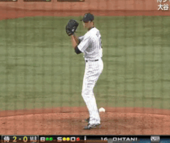
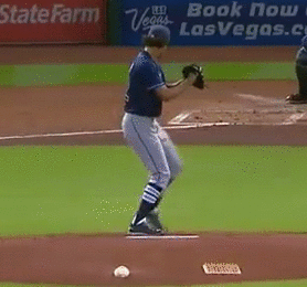
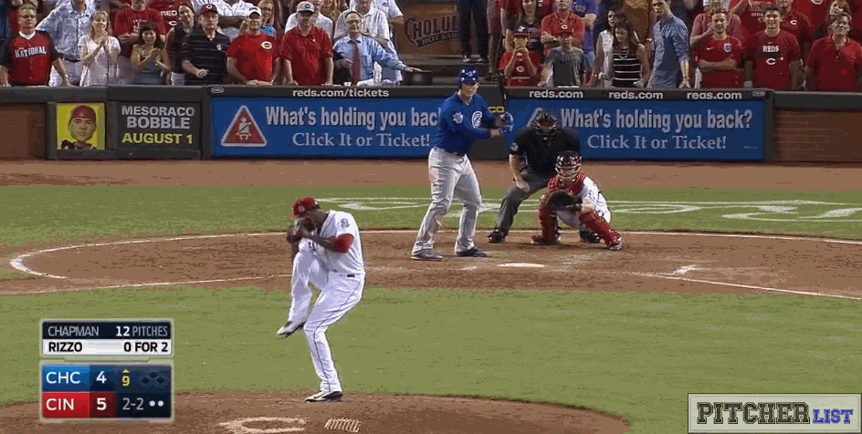
What you are observing is called dorsiflexion. Dorsiflexion is the motion about the tibiotalar (common ankle) joint which brings the foot closer towards the tibia. In all of these examples, the pitchers are accumulating potential energy prior to foot stride, by loading themselves into dorsiflexion – possibly providing them a greater ability to transfer force into kinetic energy as they move into plantarflexion and eversion (pushing down the mound towards the plate).
Now imagine one of these guys trying to throw without loading themselves into dorsiflexion first. Do you think the outcome would be the same? Would they still be able to throw with the same velocity?
Maybe.
But then again, maybe not.
Not every pitcher loads themselves in this manner prior to delivering a pitch. Having said that, a general lack of ability to dynamically load the ankle joint into dorsiflexion is something that we can screen for with our athletes.
This issue is compounded as a positional player (think: when you are fielding the ball or running), or in the weight room where several exercises require the ability for both ankles to control load through dorsiflexion (the squat, counter-movement jump, and hex-bar deadlift come to mind). If you cannot maintain and control dorsiflexion with these movements, you not only run the risk of poor quality form, you also risk injury.
Be it on the mound while pitching, or in the weight room while squatting, there are several reasons for identifying potential dorsiflexion discrepancies.
What follows is a quick update on dorsiflexion literature, and how to run a quick screen on your athletes that you think could benefit from this.
How to assess ankle dorsiflexion: a walk-through guide
As mentioned above, adequate range of motion in dorsiflexion is essential to efficient performance of fundamental weight-bearing tasks such as squatting (in our examples this could be the passive loading prior to stride) or landing movements. Landing or cutting tasks in sporting activities place high demand on structures of the ankle joint. Dorsiflexion range of motion used during each landing must absorb and dissipate the high ground reaction forces that occur upon interacting with the ground. Landing movements typically involve high impact velocities, ranging from 2.0 to more than 6.0 metres/second, which result in average peak ground reaction force of between 2.6 and 18 times body weight. As such, landing movements require rapid deceleration of the body’s momentum as the entire lower limb, beginning with the ankle joint, acts in a coordinated multi-joint fashion.
Why should you care? Reduced dorsiflexion may be a modifiable risk factor for lower extremity injury that can be easily identified during clinical examination. It’s a quick protocol, that can be highly efficient at teasing out dysfunctions.
Restricted ankle dorsiflexion range of motion can be a contributing factor in a range of ankle and knee injuries including achilles tendinitis and tendinopathy, patellar tendon tendinopathy, calf muscle tightness leading to muscle strains, anterior cruciate ligament injury, and plantar fasciitis. Weight-bearing measures are thought to more accurately reflect the available ROM during functional activities such as walking, running, or stair ambulation, and may be more reliable than measures obtained in a non-weight-bearing position.
The closed chain knee to wall or weight bearing lunge test (WBLT) method: This position places the ankle in maximal dorsiflexion, and the distance from the great toe to the wall is measured in centimeters (each centimeter corresponding to approximately 3.6° of ankle dorsiflexion). This method is inexpensive, can be performed in a variety of settings, and does not require the technical proficiency associated with a goniometer or inclinometer. The WBLT is hypothesized to be more sensitive to change compared to measures of motion in degrees, and has been shown to be more reliable than the goniometer version
Patients are instructed to:
- Remove shoes and socks.
- Face wall with the second toe, center of the heel, and knee perpendicular to wall and aligned.
- Opposite limb positioned ≈ 1 foot length behind the test foot in a tandem stance with hands on the wall (Some studies suggest that only 2 fingers from each hand is allowed to touch the wall. The reality is to just be consistent with it.)
- Perform a lunge in which the lead knee is flexed with the goal of making contact between the anterior knee and the wall while keeping the lead heel firmly planted on the floor.
- Subjects progressed in 1 cm increments until the first lunge that heel and knee contact cannot be maintained.
- Foot placement is adjusted back and forth in smaller increments (0.5cm) to achieve maximum distance from the wall while maintaining knee and heel contact.
An important question you might be asking yourself is, “Is there a normative range of asymmetry?” The majority of healthy adults exhibited lunge distance asymmetries on the WBLT of 1.5 cm or less, with no limb bias present in the observed asymmetries. These findings support the recommendation of a 2 cm or greater lunge distance asymmetry on the WBLT to delineate subjects with clinically relevant impairments.
Do you work with a pitcher who might have a dorsiflexion deficit? Give this quick screen a go, and if you have positive findings, refer to a therapist who is able to help.
References:
- Venturini, C., Ituassu, N. T., Teixeira, L. M., & Deus, C. V. O. (2006). Intrarater and interrater reliability of two methods for measuring the active range of motion for ankle dorsiflexion in healthy subjects. Brazilian Journal of Physical Therapy, 10(4), 407-411.
- Hoch, M. C., & McKeon, P. O. (2011). Normative range of weight-bearing lunge test performance asymmetry in healthy adults. Manual therapy, 16(5), 516.
- Lundgren, L. E., Tran, T. T., Nimphius, S., Raymond, E., Secomb, J. L., Farley, O. R., … & Sheppard, J. M. (2015). Development and evaluation of a simple, multifactorial model based on landing performance to indicate injury risk in surfing athletes. International journal of sports physiology and performance, 10(8), 1029-1035.
- Gabbe, B. J., Finch, C. F., Wajswelner, H., & Bennell, K. L. (2004). Predictors of lower extremity injuries at the community level of Australian football. Clinical journal of sport medicine, 14(2), 56-63.
- Malliaras, P., Cook, J. L., & Kent, P. (2006). Reduced ankle dorsiflexion range may increase the risk of patellar tendon injury among volleyball players. Journal of science and medicine in sport, 9(4), 304-309.
- Konor, M. M., Morton, S., Eckerson, J. M., & Grindstaff, T. L. (2012). Reliability of three measures of ankle dorsiflexion range of motion. International journal of sports physical therapy, 7(3), 279.
- Meir, R. A., James, Z., Whitting, J. W., & Coetzee, S. O. N. J. A. (2017). Foot to shank ratio: Does it influence ankle dorsiflexion range of motion in the knee-to-wall assessment technique?. Best of British-hiGhliGhts froM the BJsM 52, 65.
- Whitting, J. W., Steele, J. R., Mcghee, D. E., & Munro, B. J. (2011). Dorsiflexion capacity affects achilles tendon loading during drop landings. Medicine & Science in Sports & Exercise, 43(4), 706-713.
I wish I had found this sooner.
Wordle is a simple yet addictive daily word-guessing game that rose to global prominence after its public release in 2021. Created by software engineer Josh Wardle as a private pastime for family, it quickly captured a wide audience for its elegant mechanics, social shareability, and low-friction design. This article examines how Wordle works, why it resonates, criticisms and counterarguments, and its broader implications for games, education, and social media.